Nuclear radiation dose to the surroundings from patients who are undergoing nuclear medicine examinations
DOI:
https://doi.org/10.7577/radopen.1196Keywords:
Nuclear medicine, radiation dose, radiation protection, radiopharmaceutical, nuclear medicine technologists, surroundings, quantitative research methodologyAbstract
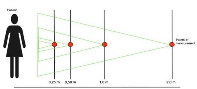
The worldwide 2009 estimates for the average annual per-capita effective radiation dose from medicine have doubled during the past 15 years. This has increased the concern for patients as radiation sources. The existing evidence indicates that the amount of radiation is small; but there are few empirical studies with results documenting the actual extent. In this study, we examined the radiation from 48 patients undergoing nuclear medical examination. 20 patients were examined with bone scintigraphy, 20 underwent MUGA, while the remaining 8 went through octreotide scintigraphy procedure. At 0.25 meters from the patient, the radiation ranged from 31±9 µSvh-1 for octreotide scintigraphy patients (111-In as agent), 69±13 µSvh-1 for bone scintigraphy (99mTc), to 92±26 µSvh-1 for the MUGA patients (99mTc). On the basis of these findings and others, one may consider current practices regarding waiting and using led shielding in areas where appropriate. Perhaps, and more important, these results could be used to improve patient and staff education. Better information material with more evidence will reduce undue anxiety.References
Aalen, O O. (1994) Introduction to statistics - with medical examples (translated from
Norwegian: Innføring i Statistikk – Med Medisinske Eksempler). Vol 2. Oslo: Ad
Notam Gyldendal
Bayram, T, Yilmaz, A H, Demir, M og Sonmez, B (2011): Radiation Dose to Technologists per Nuclear Medicine Examination and Estimation of Annual Dose, Journal of Nuclear Medicine Technology, 39, pp 55-59.
Great Norwegian encyclopedia.(2007). Disintegration. Downloaded 23.05.13, from: http://snl.no/desintegrasjon
Great Norwegian encyclopedia. (2013). Indium. Downloaded 16.05.13, from:
http://snl.no/indium
ICRP. (2000). Annual Report – Pregnancy and medical irradiation. Publication 84. Konishi E, Abe K, Kusama T (1994): Urinary excretion and external radiation dose from patients administered with Radiopharmaceuticals. Journal of Radiation protection
Dosimetry, Vol 54, No. 1, pp.61-64
Mettler et al (2009): Radiological and Nuclear medicine Studies in the United States and
Worldwide: frequency, radiation Dose, and Comparison with other Radiation Sources – 1950-2007. Journal of Radiology: Vol 253, No 2 (2009)
Leaflet Octreoscan™ from Mallinckrodt Pharmaceuticals, (01.06.2005).
Leaflet TechneScan™ HDP from Mallinckrodt Pharmaceuticals, (24.07.2007).
Leaflet TechneScan™ PYP from Mallinckrodt Pharmaceuticals, (20.12.1999).
Nuclear medicine dpt. St. Olavs Hospital, Trondheim.(2013). Procedure Bone Scintigraphy
Nuclear medicine dpt. St. Olavs Hospital, Trondheim.(2013). Procedure MUGA
Nuclear medicine dpt. St. Olavs Hospital, Trondheim.(2013). Procedure Octreotidscintigrafi
Norsk Lovtidend. (2013). Radiation Protection regulations (Norwegian: Strålevernforskriften - Norsk Lovtidend)
Downloaded 01:05.13, from http://www.lovdata.no/cgiwift/ldles?doc=/sf/sf/sf-20031121-1362.html
Piwowarska-Bilska H et al (2011): Occupational exposure at the Department of Nuclear Medicine as a work environment: A 19-year follow-up. Pol J Radiol. 2011 Apr-Jun; 76(2): 18–21.
Rootwelt, K. (2005). Nukleærmedisin 2. Utg. Oslo: Gyldendal Akademisk.
St. Olavs Hospital – MUGA (2012) Downloaded: 28.11.12, from:
http://www.stolav.no/no/Pasient/Undersokelse/Nuklearmedisinskeundersokelser/test/95527/
St. Olavs Hospital – Octreotidscintigrafi (2012) Downloaded 28.11.12, from: http://www.stolav.no/no/Pasient/Undersokelse/Nuklearmedisinskeundersokelser/Octreotidscintigrafi/96635/
St. Olavs Hospital. (2012). (Eng: bone scintigraphy) Skjelettscintigrafi med/uten innløpsfase.
Downloaded 28.11.12, from:
http://www.stolav.no/no/Pasient/Undersokelse/Nuklearmedisinskeundersokelser/Skjelettscintigrafi/95506/
The Norwegian Radiation Protection Authority. (2012). Nuclear Medicine. Downloaded
28.11.12, from: http://www.nrpa.no/nukleaermedisin
The Norwegian Radiation Protection Authority. (2012). Riation doses and limits (translated from Norwegian: Stråledoser og Grenseverdier – Statens Strålevern). Downloaded 7.12.12, from: http://www.nrpa.no/eway/default.aspx?pid=239&trg=Center_6304&CenterAndRight_6254=6304:0:15,4970:1:0:0:::0:0&Center_6304=6312:80096::1:6322:3:::0:0
The Norwegian Radiation Protection Authority. (2018). Veileder 10 – om nukleærmedisin – Downloaded 03.05.13, from http://www.nrpa.no/dav/6a19fd74c1.pdf
US EPA. (2013). Technetium-99| Radiation Protection| Downloaded:16.05.13, from http://www.epa.gov/radiation/radionuclides/technetium.html
User Manual Electronic personal dosimeter: EPD MK2+ fra Thermo Scientific
User Manual Handheld dose meter: RadEye B20 fra Thermo Scientific
Downloads
Published
2014-11-29
How to Cite
Stenstad, L.-I., Pedersen, G. A., Dypvik Landmark, A., & Brattheim, B. (2014). Nuclear radiation dose to the surroundings from patients who are undergoing nuclear medicine examinations. Radiography Open, 1(1), 7. https://doi.org/10.7577/radopen.1196
Issue
Section
Articles
License
Authors who publish with this journal agree to the following terms:- Authors retain copyright and grant the journal right of first publication, with the work after publication simultaneously licensed under a Creative Commons Attribution License that allows others to share the work with an acknowledgement of the work's authorship and initial publication in this journal.
- Authors are able to enter into separate, additional contractual arrangements for the non-exclusive distribution of the journal's published version of the work (e.g., post it to an institutional repository or publish it in a book), with an acknowledgement of its initial publication in this journal.
- Authors are permitted and encouraged to post their work online (e.g., in institutional repositories or on their website) prior to and during the submission process, as it can lead to productive exchanges, as well as earlier and greater citation of published work (See The Effect of Open Access).
Cited by


